Nerve06/eyes and ears/introduction of eye anatomy

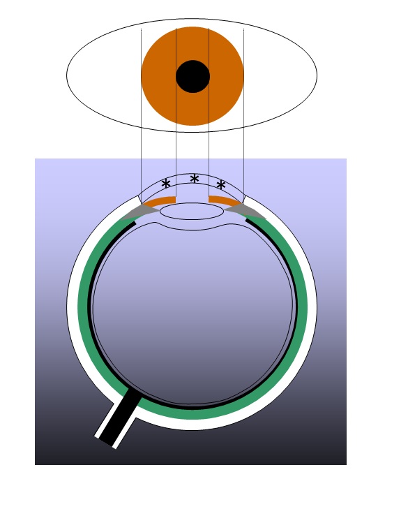
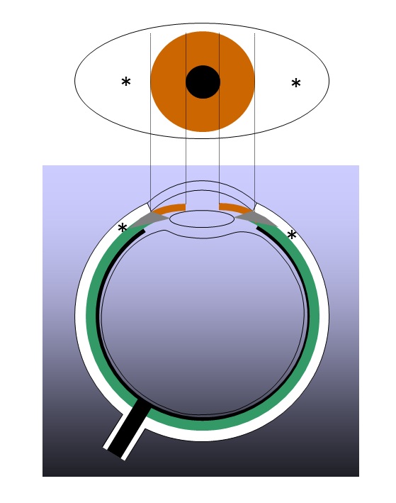
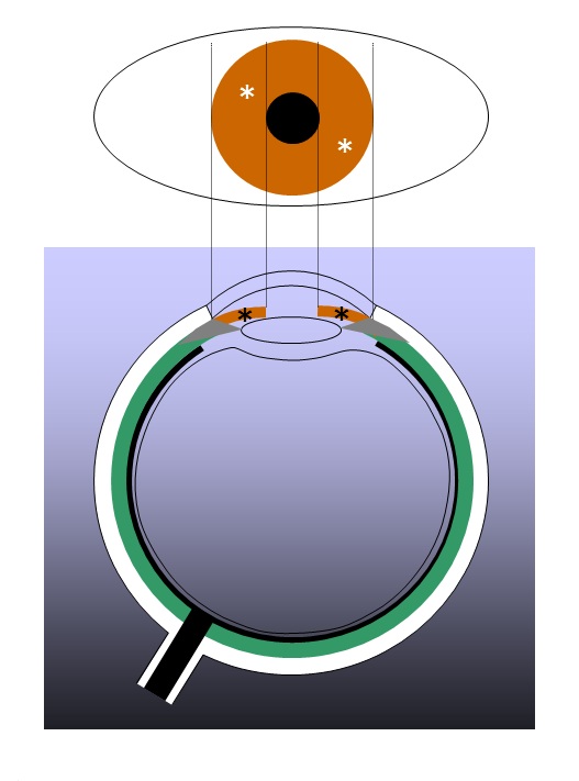
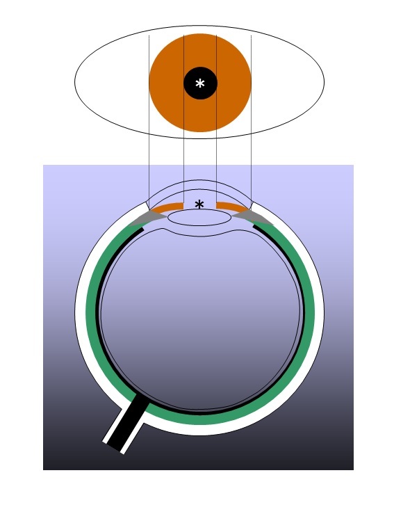
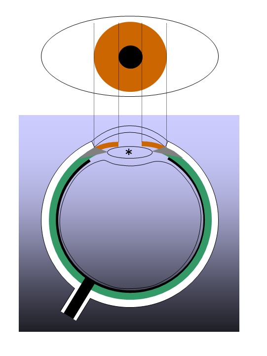
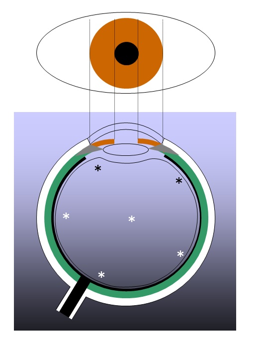
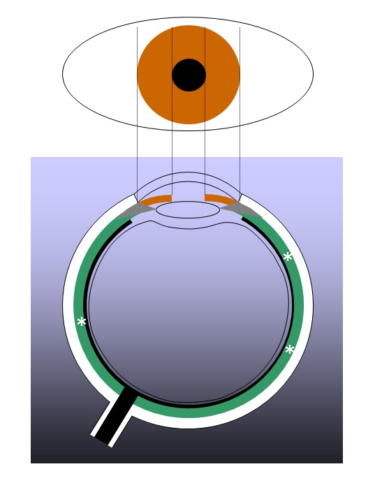
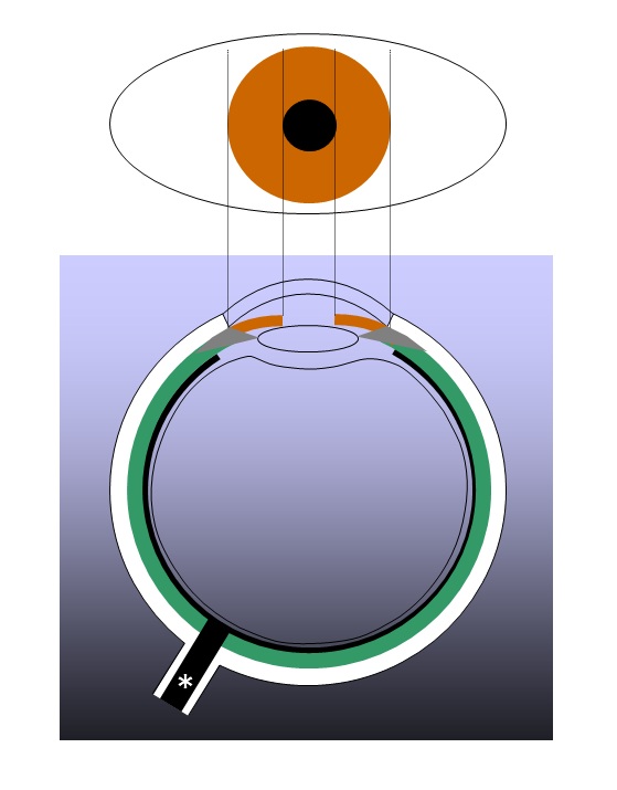
The upper half is the view from the front, and the lower half is the horizontal cross section.
The cornea is in the center of the very front plane. It is transparent, letting light into the eyes. (In the lower half, the light to dark blue gradation area is transparent).
The so-called 'white' of the eye is the conjunctiva. As with the cornea, it is in the very front plane, but is not transparent, but white, not allowing light through.
The colored area of the eye is the iris (3). It is doughnut-shaped, and changes shape to adjust the amount of light entering the eye. The hole part of the doughnut is the pupil (4). It is the name of the hole and there is nothing there. It is the entrance gate for light to enter the eye. Light passing through the pupil travels through the lens (5) which bends the light and plays a role in focusing like the lens in a camera. This is also transparent. Light, then passes through the vitreous body (6). Naturally, it is transparent. It is located at the center of the eye, forming its ball shape. Since it does not bend the light, it is not involved in focusing.
The retina (7 in green), which is just in front of the back wall of the eye, is the receptor of the light and generates an action potential.
The generated electrical signal (action potential) travels through the nerve fibers (thick black line) on the inner surface of the retina. The fibers are then bundled into one nerve.
The sensory (afferent) peripheral nerve thus formed, the optic nerve (8), travels backwards through the retina, and delivers the electric signals (action potential) to the brain.
Challenge Quiz
The asterisks indicate cornea conjunctiva iris pupil lens vitreous body retina optic nerve of the eye.
The asterisks indicate cornea conjunctiva iris pupil lens vitreous body retina optic nerve of the eye.
The asterisks indicate cornea conjunctiva iris pupil lens vitreous body retina optic nerve of the eye.
The asterisk indicates cornea conjunctiva iris pupil lens vitreous body retina optic nerve of the eye.
The asterisk indicates cornea conjunctiva iris pupil lens vitreous body retina optic nerve of the eye.
The asterisks indicate cornea conjunctiva iris pupil lens vitreous body retina optic nerve of the eye.
The asterisks indicate cornea conjunctiva iris pupil lens vitreous body retina optic nerve of the eye.
The asterisk indicates cornea conjunctiva iris pupil lens vitreous body retina optic nerve of the eye.
The part of the eye involved in adjusting the amount of light entering the eye is the cornea conjunctiva iris lens vitreous body retina optic nerve .