Nerve06/eyes and ears/introduction of eye anatomy

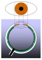
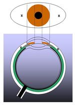
The upper half is the view from the front, and the lower half is the horizontal cross section.
The cornea is in the center of the very front plane. It is transparent, letting light into the eyes. (In the lower half, the part where the light-dark blue of the back can be seen is transparent).
The so-called 'whites' of the eye is the conjunctiva. As with the cornea, it is in the very fron plane, but is not transparent, but white, not allowing lights through.
いわゆる「茶目」の部位が「虹彩(3)」です。ドーナッツ状であり、大きさが変化することで目の中に入る光量を調節しています。
ドーナッツの穴の部分が「瞳孔(4)」です。名前の通り、穴であり、何もないのです。光が目の中に入る場所です。いわゆる「黒目」の部分です。
瞳孔を通った光は「水晶体(5)」を通ります。光を曲げる作用があり、カメラのレンズのようにピント(フォーカス・遠近)調節をしています。やはり透明です。
光は次に「ガラス体(6)」を通ります。当然、透明です。目の中心部分にあり、球状を保つ作用があります。光を曲げる作用はないので、遠近調節の作用はありません。
眼球の後壁の内側にある「網膜(7、緑色の部位)」が光の受容器であり、電気信号(活動電位)が発生します。発生した電気信号(活動電位)は網膜の内側部分にある神経線維(太い黒線)を通って、1ヶ所に集められます。
神経線維が集まってできた視神経(8)が網膜を貫いて、感覚性(求心性)末梢神経となり、脳へ電気信号(活動電位)を移動させます。
Challenge Quiz
質問 正解. 誤答. 誤答 。
この心電図の波は P. Q. R.S. T 波である。
The ECG wave in this illustration is the P. Q. R.S. T wave.
図の*印は 角膜.結膜. 虹彩. 瞳孔. 水晶体. ガラス体. 網膜. 視神経 である。
図の*印は 角膜 結膜 虹彩 瞳孔 水晶体 ガラス体 網膜 視神経 である。
眼球内にあってレンズとして作用している部位は、 胸膜 結膜 角膜 脈絡膜 網膜 毛様体 虹彩 水晶体 ガラス体 である。
眼球内にあって光量調節を担っている部位は、 胸膜 結膜 角膜 脈絡膜 網膜 毛様体 虹彩 水晶体 ガラス体 である。